
Dorsal thalamus Braininteratlas
The Stria medullaris (SM) Thalami is a discrete white matter tract that directly connects frontolimbic areas to the habenula, allowing the forebrain to influence midbrain monoaminergic output. Habenular dysfunction has been shown in various neuropsychiatric conditions.

The dorsal diencephalic conduction system, with the stria medullaris,... Download Scientific
The septal area also projects to the habenula nuclei via the stria medullaris thalami and the anterior hypothalamus. Hypothalamus. The hypothalamus lies at the center of the limbic system and is at the confluence of many neural pathways. It is subdivided from anterior to posterior into three zones: the supraoptic region, the tuberal region and.
Thalamus Earth's Lab
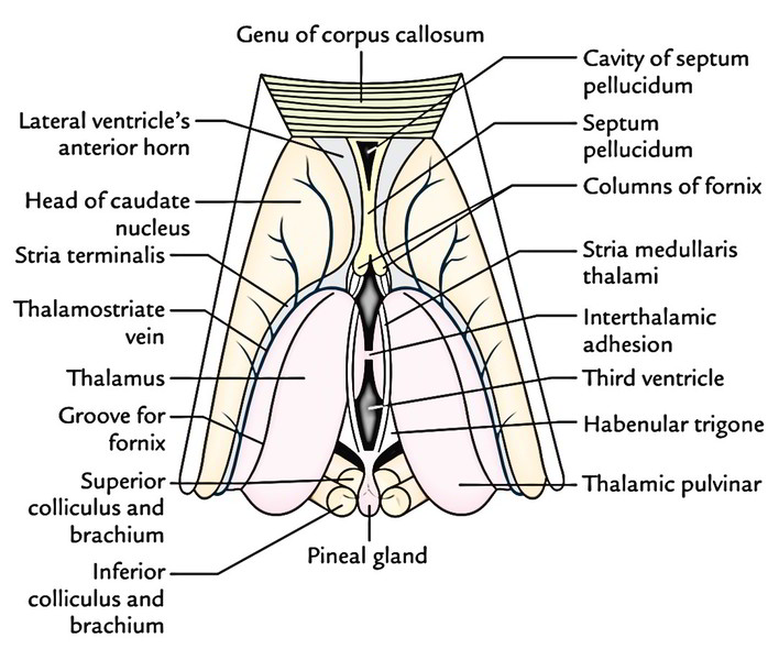
The main afferent system is composed by olfactory fibers that run within the stria medullaris thalami. The habenular complex receives these fibers, which are derived from the septal region, hypothalamus, and the amygdala. The stria medullaris thalami is a band of fibers arching on the upper part of the medial surface of the thalamus, passing.
(A) Transverse brainstem section. Stm stria medullaris; VIIIh main... Download Scientific Diagram
This nucleus communicates with the rest of the limbic system via the stria medullaris thalami (along the midline of the roof of the third ventricle). Habenula 1/2. Synonyms: none. In addition to connecting the Habenular nucleus to the hypothalamus, it also connects it to nuclei of the septum (septal area).
Frontiers Awakening Neuropsychiatric Research Into the Stria Medullaris Development of a
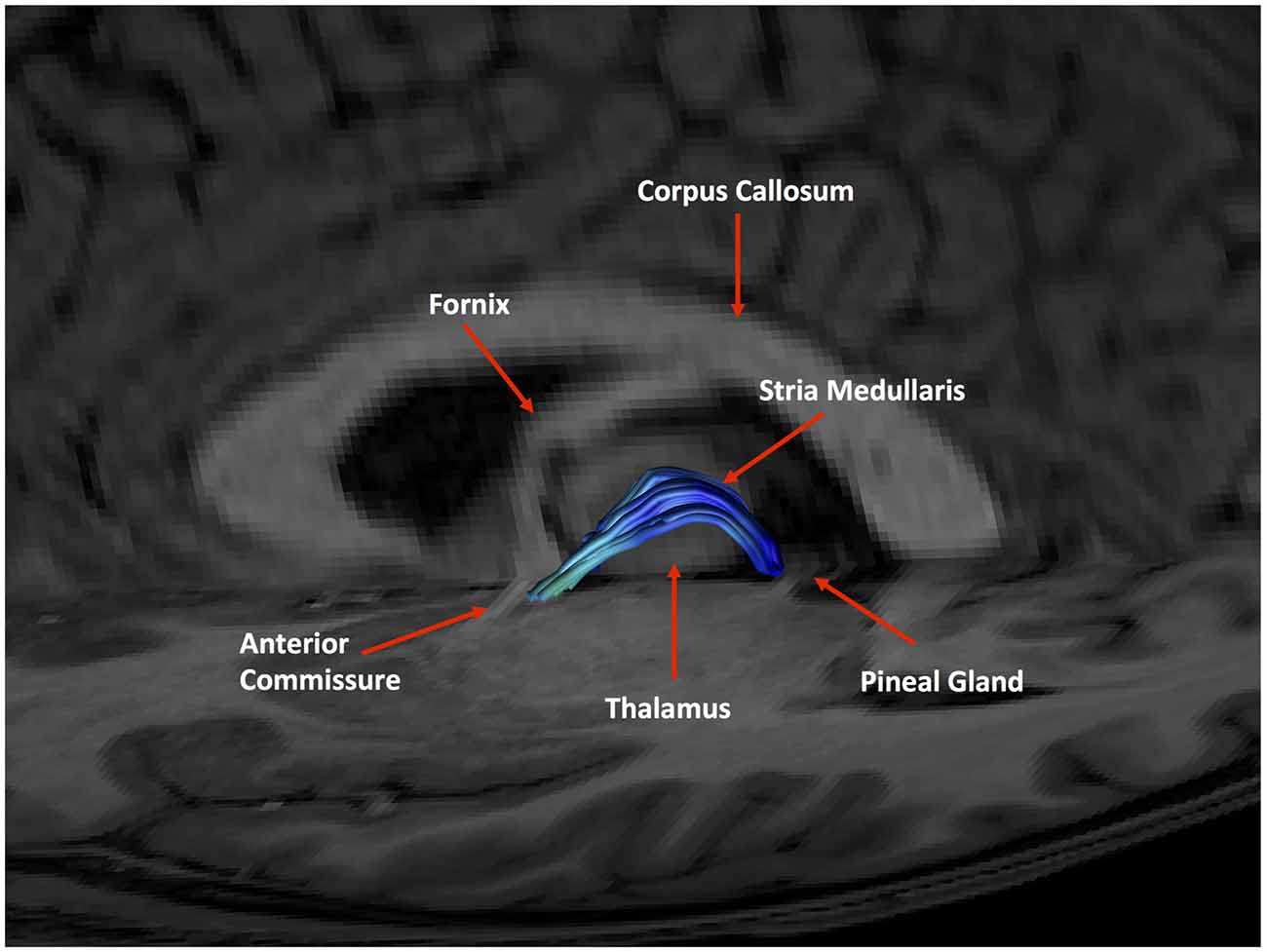
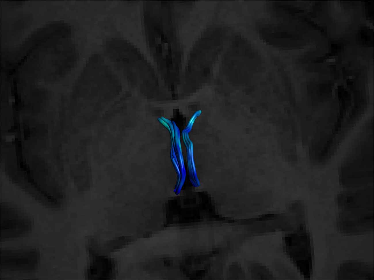
Abstract Background: Little is known of its significance, especially in regard to functional pathways. Probabilistic diffusion tensor imaging (DTI) has recently been used to seed the lateral habenula and define its afferent white matter pathway, the stria medullaris thalami (SM).
Frontiers Awakening Neuropsychiatric Research Into the Stria Medullaris Development of a
A bundle of fibers called the stria medullaris thalami are located near the junction of medial and superior (upper) surfaces. Lateral Surface. The lateral surface of the thalamus is covered by a layer of myelinated fibers called the external medullary lamina which separates the lateral surface from the reticular nuclei.

727 Graustufen Stria Medullaris Stockfotografie Alamy
It is the caudal part of the forebrain (prosencephalon) that occupies the central region of the brain. The diencephalon is comprised of the: Epithalamus Thalamus Subthalamus Metathalamus Hypothalamus In the following article, we will explore the anatomy of different parts of the diencephalon as well as their function. Contents Function
PPT Thalamus PowerPoint Presentation, free download ID9508009
The Stria medullaris (SM) Thalami is a discrete white matter tract that directly connects frontolimbic areas to the habenula, allowing the forebrain to influence midbrain monoaminergic output. Habenular dysfunction has been shown in various neuropsychiatric conditions.
Dorsal thalamus Braininteratlas
Stria medullaris thalami - e-Anatomy - IMAIOS Human anatomy 2 Human body Parts of human body Regions of human body Musculoskeletal systems Visceral systems Integrating systems Endocrine glands Cardiovascular system Lymphoid organs Nervous system Central nervous system Gray matter White matter Reticular formation Ependyma Meninges Brain Cerebrum
stria medullaris 4th ventricle
Recent literature has implicated the cortico-striatal-pallidal-thalamic loop in the pathophysiology of MDD which implies that stimulation of an afferent white matter tract like the SM modulates a node within a broad network involved in the pathophysiology of mood disorders ( Downar et al., 2016
Frontiers Awakening Neuropsychiatric Research Into the Stria Medullaris Development of a
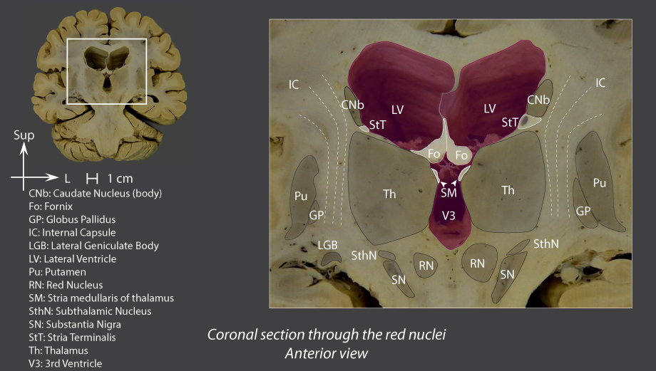
The habenular nuclei receive afferents via the stria medullaris thalami from the septal area (subcallosal area) and pre-optic nuclei (hypothalamus), via stria terminalis from the amygdaloid body and via fornix from the hippocampus (1, 4-8, 25-28). Some stria medullaris thalami fibers pass through the dorsal lamina of the pineal stalk and.

Thalamus Anatomy
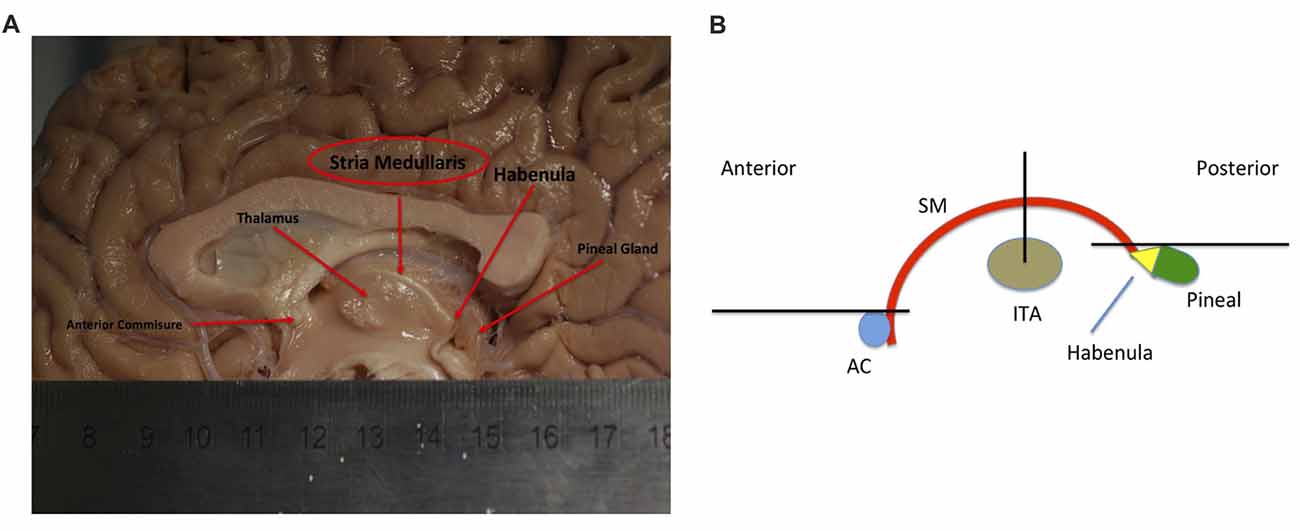
The stria medullaris (SM), (Latin, furrow and pith or marrow) is a part of the epithalamus and forms a bilateral white matter tract of the initial segment of the dorsal diencephalic conduction system (DDCS). It contains afferent fibers from the septal nuclei, lateral preoptico- hypothalamic region, and anterior thalamic nuclei to the habenula.

DIENCEPHALON David Kachlík Petr Zach diencephalon
Identification of the stria medullaris thalami using diffusion tensor imaging Identification of the stria medullaris thalami using diffusion tensor imaging Neuroimage Clin. 2016 Oct 26;12:852-857. doi: 10.1016/j.nicl.2016.10.018. eCollection 2016. Authors Ryan B Kochanski 1 , Robert Dawe 2 , Daniel B Eddelman 1 , Mehmet Kocak 3 , Sepehr Sani 1

PPT Subthalamus & Hypothalamus PowerPoint Presentation ID1159156
Stria medullaris thalami; Locate closely to the taenia thalami is the stria medullaris, a group of fibers that run along the joint of the superior and medial parts of the thalamus. The stria medullaris runs along the length of the anterior pole of the thalamus towards the Habenular trigone.
Thalamic Nuclei Connections, Functions & Anatomy Kenhub
It lies most medially and adjacent to the AV but is separated from AV, the stria medullaris thalami, and the ventricular surface of myelinated fibers and a glial lamella. Our results show that the.
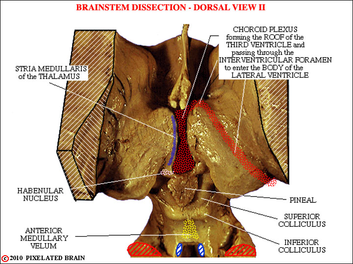
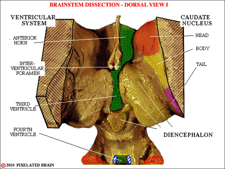
Pixelated Brain Module 2, Section 2 Dorsal views of the brainstem
Awakening Neuropsychiatric Research Into the Stria Medullaris: Development of a Diffusion-Weighted Imaging Tractography Protocol of This Key Limbic Structure Front Neuroanat. 2018 May 8;12:39. doi: 10.3389/fnana.2018.00039. eCollection 2018. Authors